Cardiomyopathy Types Explained: Dilated, Hypertrophic, and Restrictive
 Jul, 3 2026
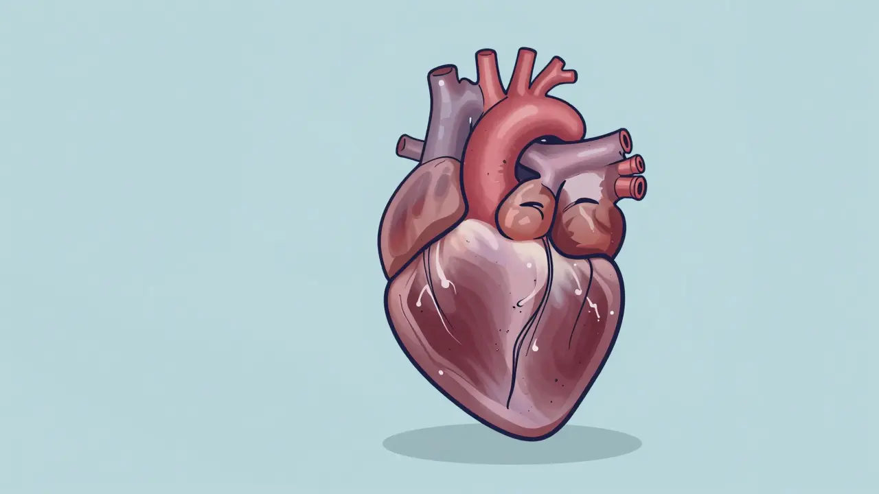
Your heart is a pump. It’s supposed to squeeze blood out and relax to fill up again. When the muscle itself gets sick, that rhythm breaks. This isn’t just about clogged arteries or high blood pressure. This is cardiomyopathy, a primary disease of the myocardium where the heart muscle becomes enlarged, thickened, or stiff, impairing its ability to pump effectively. If you’ve heard this term, it probably sounded like a vague label for "bad heart." But in reality, it’s a specific group of conditions with very different causes, symptoms, and treatments. Getting the type right is the difference between taking the wrong medication and getting your life back on track.
We often lump heart problems together, but cardiomyopathy is distinct from coronary artery disease. While a heart attack kills muscle due to lack of oxygen, cardiomyopathy changes the structure of the muscle itself. According to data from the National Institutes of Health, these three main types-dilated, hypertrophic, and restrictive-account for about 90% of all cases. Understanding which one you’re dealing with requires looking at what the heart looks like on an echo and how it behaves under stress.
Dilated Cardiomyopathy: The Stretched Heart
Imagine blowing up a balloon until the rubber gets thin and floppy. That’s essentially what happens in Dilated Cardiomyopathy (DCM). The left ventricle-the main pumping chamber-stretches out and enlarges. Because the walls are stretched thin, they can’t squeeze hard enough to push blood forward.
This is the most common form, representing 50-60% of all cardiomyopathy cases. You might not feel anything at first, but as the heart struggles, fluid backs up into your lungs or legs. Common signs include shortness of breath when lying flat, swelling in the ankles, and extreme fatigue. Your doctor will look for an ejection fraction below 40%. Normal hearts pump out 50-70% of their blood with each beat; DCM hearts struggle to hit half that number.
Why does this happen? Sometimes it’s genetic, involving mutations in genes like TTN or LMNA. Other times, it’s acquired. Chronic heavy alcohol use, certain chemotherapy drugs like doxorubicin, or viral infections like coxsackievirus can trigger it. In some cases, we never find a cause-it’s called idiopathic. The good news? Many people see significant improvement with guideline-directed medical therapy. Drugs like sacubitril/valsartan have shown a 20% greater reduction in heart failure markers compared to older medications, helping some hearts actually shrink back toward normal size.
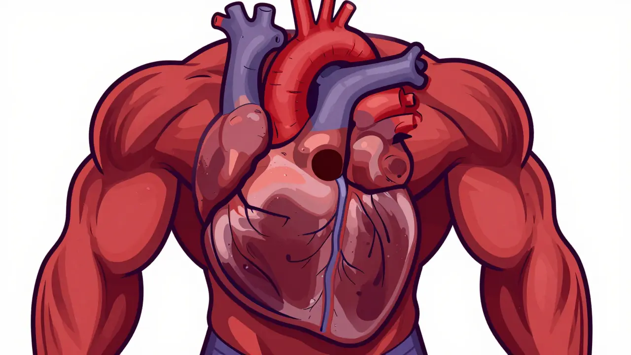
Hypertrophic Cardiomyopathy: The Thickened Heart
If DCM is a stretched balloon, Hypertrophic Cardiomyopathy (HCM) is a heart that has worked out too much. The muscle walls become abnormally thick, often without any obvious reason like high blood pressure.
This condition affects roughly 1 in 500 people. It’s famously known as the leading cause of sudden cardiac death in young athletes under 35. The thickening usually happens in the septum-the wall between the two lower chambers. This can block blood flow out of the heart, creating a gradient that makes every heartbeat harder work. On an echocardiogram, doctors look for a wall thickness of 15 mm or more in adults.
HCM is overwhelmingly genetic. About 60% of cases involve mutations in sarcomere proteins like MYH7 or MYBPC3. It runs in families, often skipping generations or showing up differently in siblings. One person might be asymptomatic, while another collapses during a game. Diagnosis often involves a combination of echo and genetic testing. A 17-gene panel can identify the culprit in many cases, though it doesn’t always predict severity. Treatment focuses on slowing the heart rate with beta-blockers to improve filling time. For severe obstruction, procedures like septal myectomy or new drugs like mavacamten can dramatically reduce symptoms.
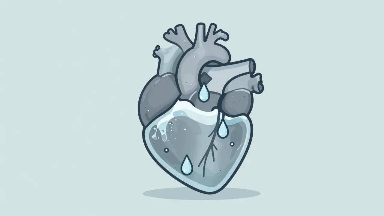
Restrictive Cardiomyopathy: The Stiff Heart
Restrictive Cardiomyopathy (RCM) is the rarest and often the most misunderstood. Here, the heart muscle isn’t necessarily weak or thick; it’s stiff. Think of trying to pour water into a rigid plastic bottle versus a flexible one. The heart can pump normally, but it can’t relax enough to fill up with blood between beats.
RCM accounts for only 5-10% of cases. The pumping strength (ejection fraction) stays normal, often above 50%, but the filling pressures skyrocket. This leads to backup in the veins, causing liver congestion, ascites (fluid in the belly), and swollen legs. The hallmark sign on an echo is a restrictive filling pattern, where blood rushes in quickly then stops abruptly because the stiff walls resist expansion.
The cause is usually an infiltrative process. Something foreign is depositing itself in the heart tissue. Amyloidosis is the big one, where misfolded proteins build up in the muscle. Sarcoidosis, hemochromatosis (iron overload), and Fabry disease are other culprits. Diagnosing RCM is tricky because it mimics constrictive pericarditis-a problem with the sac around the heart rather than the muscle itself. Cardiac MRI is crucial here, looking for late gadolinium enhancement patterns that signal fibrosis or infiltration. Treatment depends entirely on the underlying cause. For amyloidosis, drugs like tafamidis can slow progression, while iron chelation helps in hemochromatosis.
How Doctors Tell Them Apart
You might wonder how clinicians distinguish these three. They don’t just guess. They use a layered approach starting with an echocardiogram, moving to cardiac MRI, and often ending with genetic testing or biopsy.
| Type | Heart Structure | Pumping Function (EF) | Primary Issue | Common Causes |
|---|---|---|---|---|
| Dilated (DCM) | Enlarged, thin walls | Reduced (<40%) | Systolic dysfunction (weak squeeze) | Viral, alcohol, genetic, idiopathic |
| Hypertrophic (HCM) | Thickened walls | Normal or High | Diastolic dysfunction + Obstruction | Genetic (sarcomere mutations) |
| Restrictive (RCM) | Normal size, stiff walls | Normal (>50%) | Severe diastolic dysfunction (poor filling) | Amyloidosis, sarcoidosis, iron overload |
Echocardiography is the first stop. It shows chamber size, wall thickness, and valve function. But echoes have limits. That’s where Cardiac Magnetic Resonance (CMR) comes in. CMR provides a detailed map of tissue composition. It can spot scar tissue (fibrosis) in DCM, quantify the exact thickness in HCM, and detect the unique infiltration patterns of amyloidosis in RCM. Genetic testing is now standard for familial cases. In HCM, finding a mutation confirms the diagnosis and prompts screening for relatives. In DCM, it helps rule out treatable metabolic causes.
Treatment Paths and Prognosis
There is no single cure for cardiomyopathy, but management has improved drastically. The goal shifts depending on the type. For DCM, we aim to reduce strain on the heart and prevent remodeling. Standard care includes ACE inhibitors, beta-blockers, and SGLT2 inhibitors. These drugs have been shown to cut mortality by 30% over three years. In advanced cases, devices like ICDs (implantable cardioverter-defibrillators) protect against sudden arrest, and LVADs (left ventricular assist devices) can bridge patients to transplant.
HCM treatment is more personalized. If there’s no obstruction, beta-blockers or calcium channel blockers help the heart relax. If there is obstruction, we add disopyramide or consider surgical septal reduction. The approval of mavacamten in 2022 marked a turning point-it’s the first drug designed specifically to reverse the molecular hypercontraction in HCM, offering relief to those who failed other therapies.
RCM remains the hardest to treat because the damage is often structural and irreversible. We focus on managing symptoms-diuretics to remove fluid-and treating the root cause. For transthyretin amyloidosis, stabilizing agents like tafamidis have extended survival significantly. However, prognosis varies widely. Five-year survival for treated DCM is 70-80%, non-obstructive HCM is near 95%, but RCM can drop to 30-50% depending on the underlying disease.
Living with Cardiomyopathy
Receiving this diagnosis feels heavy. But it’s not a static sentence. Many people live full lives with careful management. Regular follow-ups are non-negotiable. You’ll need periodic echoes to monitor changes. Lifestyle adjustments matter too. Limiting sodium helps control fluid retention. Avoiding excessive alcohol is critical, especially for DCM. For HCM patients, competitive sports may be restricted due to arrhythmia risk, but moderate activity is generally encouraged.
Family screening is vital. Since genetics play such a large role, your siblings and children should be evaluated. Early detection in relatives can prevent sudden events. Joining support groups, like those hosted by the Cardiomyopathy Foundation, connects you with others navigating the same path. They share practical tips on diet, exercise, and navigating insurance for expensive therapies.
Is cardiomyopathy hereditary?
Yes, many forms are. Hypertrophic cardiomyopathy is predominantly genetic, with autosomal dominant inheritance in 90% of familial cases. Dilated cardiomyopathy has a genetic component in 25-35% of cases. Restrictive cardiomyopathy is less commonly inherited directly but can result from genetic storage diseases like Fabry disease. If you have cardiomyopathy, first-degree relatives should undergo screening.
Can cardiomyopathy be reversed?
In some cases, yes. Dilated cardiomyopathy caused by reversible factors like alcohol abuse, thyroid issues, or certain toxins can improve or even resolve with treatment. Medications like ARNIs and SGLT2 inhibitors can also lead to reverse remodeling, where the heart shrinks back toward normal size and function. However, hypertrophic and restrictive types are generally progressive and managed rather than cured.
What is the difference between heart failure and cardiomyopathy?
Cardiomyopathy is a disease of the heart muscle itself. Heart failure is a clinical syndrome where the heart can't pump enough blood to meet the body's needs. Cardiomyopathy is a leading cause of heart failure, but heart failure can also result from high blood pressure, valve disease, or coronary artery disease without primary muscle involvement.
How is restrictive cardiomyopathy diagnosed?
Diagnosis is challenging because symptoms mimic other conditions. Doctors use echocardiography to show normal pumping but poor filling. Cardiac MRI is essential to detect infiltration patterns like amyloidosis. In some cases, an endomyocardial biopsy is needed to confirm the specific type of tissue deposition, such as amyloid or iron.
Are there new treatments for cardiomyopathy in 2026?
Yes, precision medicine is advancing rapidly. Mavacamten is now standard for obstructive HCM. Tafamidis is widely used for ATTR amyloidosis. Gene therapies targeting specific mutations in HCM are entering clinical trials. Additionally, polygenic risk scoring is becoming available to predict susceptibility before symptoms appear, allowing for earlier monitoring.