Pulmonary Embolism: Recognizing Sudden Shortness of Breath and Diagnosis
 Apr, 9 2026
Imagine sitting on your couch watching TV when, without any warning, you suddenly feel like you can't get enough air. It isn't a slow build-up of fatigue; it's an abrupt, terrifying sensation of suffocation. This is the hallmark of a pulmonary embolism is a life-threatening condition where a blood clot blocks one or more arteries in the lungs, stopping blood flow and oxygen exchange. Because it often mimics less severe issues like anxiety or a mild chest cold, many people dismiss the warning signs until it's almost too late.
Quick Summary of Key Facts
- Primary Symptom: Sudden shortness of breath occurs in 85% of cases.
- Common Origin: About 70% of clots start as Deep Vein Thrombosis (DVT) in the legs.
- Gold Standard Test: CT Pulmonary Angiography (CTPA) is the most accurate diagnostic tool.
- Critical Warning: Pleuritic chest pain (sharp pain when breathing) is a major red flag.
Why It Happens: The Journey of a Clot
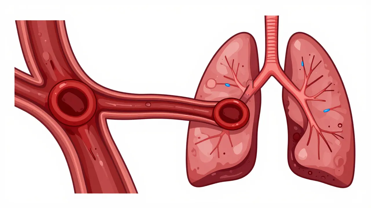
A pulmonary embolism doesn't just appear in the lungs out of nowhere. In the vast majority of cases, it starts as a Deep Vein Thrombosis is a condition where a blood clot forms in a deep vein, usually in the legs. When a piece of this clot breaks off, it travels through the bloodstream, passes through the heart, and eventually wedges itself in the pulmonary arteries. Once the pipe is blocked, the lung tissue doesn't get the blood it needs to pick up oxygen, leading to a rapid drop in blood oxygen levels.
While leg clots are the usual suspects, some people develop clots in the upper limbs, though this is rare and typically happens after the placement of a venous catheter. Whether the clot is small and peripheral or large and central, the result is the same: your body is fighting for air while your heart struggles to pump blood against a blockage.
Spotting the Red Flags: More Than Just Breathlessness
The danger of a PE is that its symptoms are "nonspecific." This means they look like a dozen other things. However, the patterns are telling. Sudden shortness of breath (dyspnea) is the big one, appearing in 85% of patients. If you are hemodynamically unstable-meaning your blood pressure is crashing-this breathlessness is often profound and occurs even while resting.
Then there is the chest pain. About 74% of people experience it, and it's usually "pleuritic," meaning it feels sharp and gets worse every time you take a deep breath or cough. This is why so many people are initially misdiagnosed with pneumonia or a heart attack. Other signs to watch for include:
- Leg Swelling: Occurs in 44% of cases, often indicating the original DVT.
- Fast Heart Rate: Tachycardia (over 100 bpm) happens in 30% of patients as the heart tries to compensate for low oxygen.
- Coughing: About 53% of people cough, and in 23% of those cases, they may cough up blood (hemoptysis).
- Fainting: Syncope happens in about 14% of cases, often signaling a massive clot that is severely restricting blood flow.
The Diagnostic Path: How Doctors Find the Clot
Because PE is so dangerous, doctors use a tiered system to rule it in or out. They don't just jump to a scan for everyone; they start by assessing your probability using scoring systems like the Wells Criteria is a clinical prediction rule used to estimate the probability of a pulmonary embolism based on patient symptoms and risk factors. If your score is low, they might use a blood test. If it's high, they move straight to imaging.
The first laboratory stop is usually the D-dimer test is a blood test that measures a substance released when a blood clot breaks down. If this test is negative (usually below 500 ng/mL), it's very effective at ruling out a PE in low-risk patients. However, it's not a perfect tool. For people over 50 or those with cancer, D-dimer levels naturally rise, which can lead to false positives. This is why age-adjusted thresholds are now used to avoid sending too many people for unnecessary radiation.
| Test | Primary Use | Sensitivity | Specificity | Key Limitation |
|---|---|---|---|---|
| CTPA | Gold Standard Confirmation | 95% | 96% | Radiation & contrast risk |
| V/Q Scan | Alternative for kidney issues | 85% | 95% | Requires nuclear facility |
| Ultrasound | Finding the source (DVT) | >90% | 95% | Doesn't see the lung clot |
| D-dimer | Ruling out (low risk) | 97% | Low in elderly | High false positives |
The Gold Standard: CTPA and Alternatives
When a doctor needs a definitive answer, they use Computed Tomography Pulmonary Angiography is a specialized CT scan that uses an iodinated contrast dye to visualize the pulmonary arteries (CTPA). This test allows them to see exactly where the blockage is. It's incredibly accurate, detecting clots in 92% of cases when using 3 mm slices.
But what if you can't have contrast dye? Some people have kidney failure or severe allergies. In those cases, doctors turn to a V/Q Scan is a nuclear medicine test that compares air flow (ventilation) and blood flow (perfusion) in the lungs. It's slightly less sensitive than a CT, but it's a lifesaver for patients who can't handle contrast. For those who are completely unstable-too sick to even be moved to a radiology suite-doctors use bedside echocardiography to look for signs of right ventricular strain, which is a strong indicator of a massive PE.

The Danger of Delay: Why Diagnosis Often Fails
The scariest part of pulmonary embolism isn't just the clot-it's the delay in finding it. Real-world data shows that many patients visit healthcare providers more than twice before getting the correct diagnosis. Some are told they have anxiety; others are told it's just asthma or pneumonia. This happens because the symptoms are so varied. You might feel fine one minute, and the next, you're gasping for air while sitting still.
These delays can be fatal. However, when hospitals implement a structured Pulmonary Embolism Response Team is a multidisciplinary team of specialists who coordinate the rapid diagnosis and treatment of PE (PERT), the results are dramatic. One health system saw the time to get a CT scan drop from 127 minutes to just 43 minutes, which slashed mortality rates from 8.2% down to 3.1%. Speed is everything when your lungs aren't getting oxygen.
Looking Forward: AI and New Markers
The way we find these clots is changing. We're seeing the rise of AI-driven interpretation, such as the PE-Flow algorithm, which helps radiologists spot clots with incredible precision. There is also a push toward better biomarkers. While the D-dimer is the current standard, researchers are testing new panels that combine D-dimer with other proteins to create a test that is nearly 99% accurate in ruling out PE for intermediate-risk patients.
If you've had a clot before, stay vigilant. About 33% of people who've had a DVT or PE will experience another one within ten years. Your risk is also significantly higher-nearly five times higher-if you are fighting cancer. Being aware of these risk factors can be the difference between a quick trip to the ER and a critical emergency.
Can a pulmonary embolism be mistaken for a panic attack?
Yes, very frequently. Both can cause sudden shortness of breath, chest tightness, and a feeling of dread. However, PE is often accompanied by physical signs like leg swelling, a very high heart rate, or a sharp pain that worsens with a deep breath, which are not typically present in a panic attack.
Is a D-dimer test enough to rule out a PE?
Only if your overall clinical risk is low. For low-risk patients, a negative D-dimer is very reliable (97% sensitivity). However, for people over 50 or those with cancer, D-dimer levels often stay elevated regardless of a clot, meaning a positive result doesn't always mean you have a PE, and a negative might not be enough to rule it out without imaging.
What is the difference between DVT and PE?
DVT (Deep Vein Thrombosis) is the "source"-a clot that forms in a deep vein, usually in the leg. A PE (Pulmonary Embolism) is the "event"-it happens when that DVT clot breaks loose, travels through the heart, and gets stuck in the lung arteries.
Why is CTPA called the gold standard?
CTPA provides high-resolution, direct visualization of the pulmonary arteries. With 95% sensitivity and 96% specificity, it is the most accurate way to see exactly where and how large a clot is, allowing doctors to make immediate treatment decisions.
What should I do if I suspect I have a pulmonary embolism?
Seek emergency medical care immediately. Because PE can lead to obstructive shock and sudden cardiac arrest, it requires immediate intervention. Do not wait to see if the shortness of breath goes away on its own.
Danny Wilks
April 9, 2026 AT 18:52It's quite fascinating how the medical community has evolved to implement these PERT teams to streamline the diagnostic pipeline, as the intersection of bureaucracy and emergency medicine often creates these tragic delays that could be easily mitigated by better organizational structures.
danny Gaming
April 10, 2026 AT 02:17Typical gov healthcare fail... we dont need fancy teams we need doctors who actually do their jobs right the first time without needing a manual lol
Trey Kauffman
April 11, 2026 AT 22:56Oh sure, because nothing says "modern medicine" like visiting the ER three times before someone notices you're actually dying of a clot. Pure genius. 🙄
Doug DeMarco
April 12, 2026 AT 17:37Glad this info is out there! Definitely worth keeping an eye on those legs after a long flight ✈️ stay safe everyone! 😊
Simon Jenkins
April 14, 2026 AT 09:09I simply cannot abide the lack of mention regarding the sheer psychological trauma of a misdiagnosis! Imagine the absolute horror of being told it's just a panic attack while your lungs are literally failing! It is an affront to the patient's dignity and a catastrophic failure of the clinical gaze! I've seen this tragedy unfold in the most opulent of clinics and it's just heart-wrenching!
Peter Meyerssen
April 14, 2026 AT 17:27The dichotomy of the D-dimer's sensitivity versus its specificity is just a metaphor for the human condition... we seek certainty in a world of biological noise 🌀. It's all about that hemodynamic instability, man. ✌️
Victor Parker
April 16, 2026 AT 09:31Ever wonder why these "gold standard" tests only became popular now? Maybe they're hiding something about how these clots actually start... looks like a way to push more expensive dyes on us 🤨
Will Gray
April 17, 2026 AT 09:20Exactly. The push for AI-driven interpretation is just the next step in the global surveillance state. First they scan your lungs for clots, then they scan your brain for dissent. Only a true patriot sees through this medical industrial complex.
Franklin Anthony
April 18, 2026 AT 10:23it is honestly just a shame how the system works but i agree the ai part is a bit sketchy if you think about the data ownership side of things
Camille Sebello
April 20, 2026 AT 07:58Leg swelling is key!!! Check your calves!!! Now!!!
Sarina Montano
April 22, 2026 AT 03:37Actually, if you're in a pinch and can't get a CTPA, the V/Q scan is a brilliant alternative for those with compromised renal function. It's a bit like a puzzle where you compare the air-map with the blood-map to find the missing piece. It's not as flashy as a high-res CT, but it's an absolute lifesaver in the right clinical context, especially when you're dealing with contrast allergies that could trigger anaphylaxis. Plus, bedside echo is a total game-changer for those too unstable to move, giving a glimpse of the heart's right ventricle struggling against that blockage in real-time. It's honestly amazing how many layers of safety we have if the doctor actually knows where to look. I've always found the hemodynamics of this fascinating because it's a race against time. The whole process from DVT to PE is like a dangerous journey through the venous highway. Once that clot hits the pulmonary artery, the pressure spikes are wild. You really have to appreciate the complexity of the pulmonary vasculature. It's a delicate balance of pressure and flow. When that balance breaks, the symptoms are a symphony of distress. From the tachypnea to the tachycardia, your body is screaming for help. The D-dimer is a great first filter, but it's far from a crystal ball. You need the full clinical picture to avoid those deadly misdiagnoses. I'm so glad we're moving toward better biomarkers because a 99% rule-out rate would save countless lives every single day. It's just a matter of implementation and training. We need more providers to recognize that pleuritic pain isn't just a bad cough. The intersection of risk factors like cancer or recent surgery makes this a high-stakes game. Stay vigilant, stay informed, and always trust your gut if you feel like you're suffocating.
Ryan Hogg
April 22, 2026 AT 15:34I read this and just felt a wave of anxiety. My uncle had this and the fear in his eyes when he couldn't breathe... it just haunts me. Every time I feel a bit winded now, I just spiral into this dark place where I'm sure it's happening to me too. It's just so heavy to think about how fragile we are.
kalpana Nepal
April 23, 2026 AT 20:46India has the best doctors who can find these things without all these expensive machines they use in the west. We trust our skills and our land.