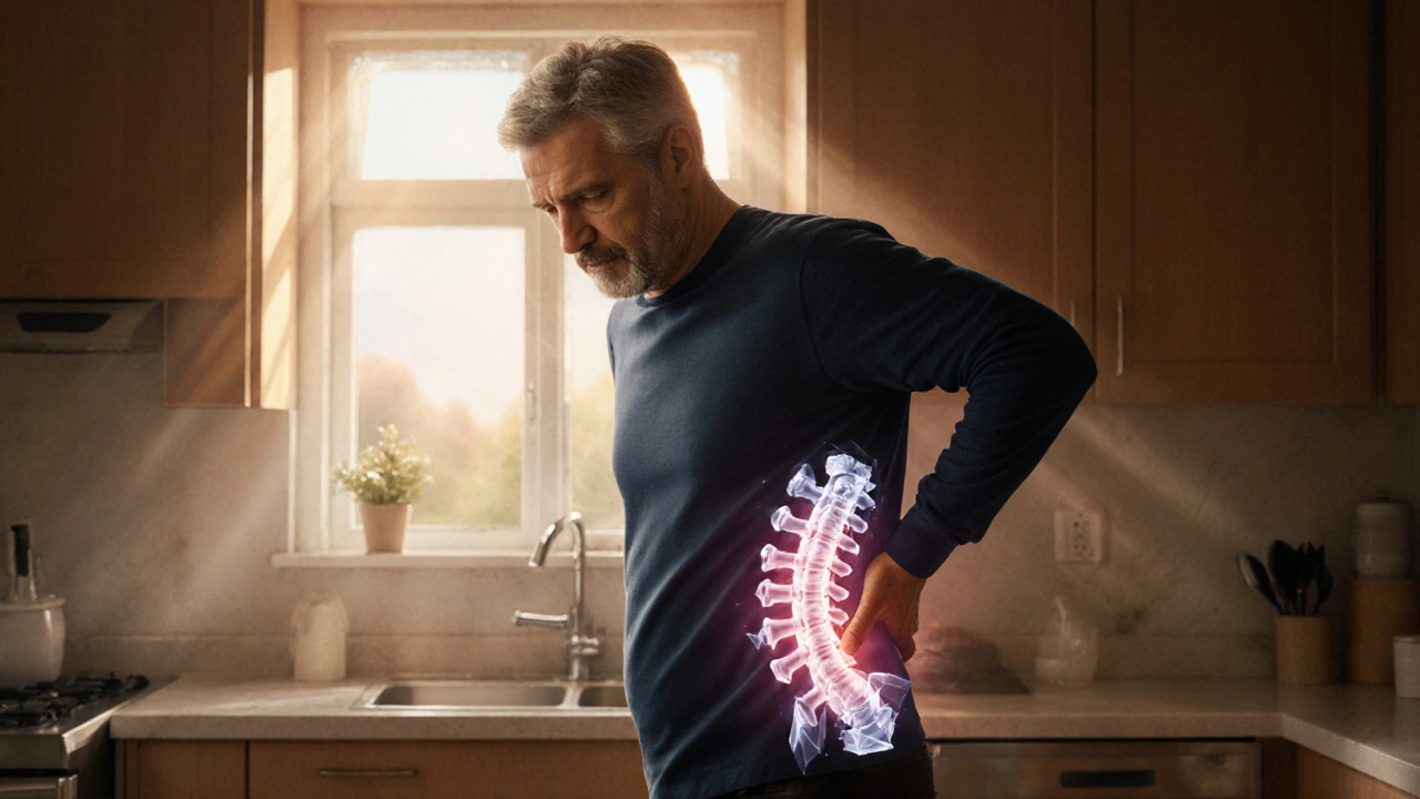
Gout’s Effect on the Spine: Symptoms, Diagnosis & Treatment
 Sep, 28 2025
Gout is a metabolic disorder where excess uric acid forms sharp crystals that lodge in joints and surrounding tissues. Most people picture swollen big toes, but the crystals can settle anywhere - even between vertebrae. When they do, the spine can become a hidden source of pain, stiffness, and nerve irritation. Understanding how gout and spine interact helps you spot red flags early, get the right imaging, and choose treatments that target the root cause instead of just masking discomfort.
TL;DR
- Spinal gout is rare but real - uric‑acid crystals can collect in facet joints, ligaments, or even the spinal canal.
- Typical signs include sudden, intense back pain with swelling, warmth, and sometimes radiating leg numbness.
- Diagnosis relies on blood uric‑acid tests plus imaging (CT or MRI) that reveals crystal deposits.
- First‑line treatment uses NSAIDs, colchicine, or steroids; long‑term control needs urate‑lowering drugs like allopurinol.
- Diet, hydration, weight management, and regular check‑ups with a rheumatologist can prevent future attacks.
How Gout Takes Hold in the Spine
The spine is a stack of vertebrae separated by intervertebral discs and supported by facet joints, ligaments, and muscles. While the discs themselves lack blood supply, the facet joints are richly vascularized - a perfect playground for uric‑acid crystals. When serum urate levels stay above 6.8mg/dL, the crystals can drift into these joints, triggering an inflammatory cascade.
Inflammation in the spine differs from peripheral joints because the spinal canal houses the spinal cord and nerve roots. A crystal‑induced swelling can compress a nerve, producing symptoms that mimic herniated discs or spinal stenosis. That’s why a single gout flare can feel like a full‑blown back injury.
Common Symptoms That Hint at Spinal Gout
Because back pain is ubiquitous, you need specific clues to suspect gout:
- Sudden onset - pain erupts within hours, often at night.
- Localized warmth and redness over the affected vertebral level.
- Sharp, throbbing pain that may radiate to the buttock or down the leg (sciatica‑like).
- Joint stiffness that improves modestly with movement but worsens after rest.
- Accompanying flare elsewhere - such as the big toe or ankle - points toward systemic urate issues.
If you’ve experienced any of these alongside a known history of gout, bring them up with your doctor.
How Doctors Diagnose Spinal Gout
Diagnosing gout in the back isn’t as simple as swabbing a joint. It combines lab work, imaging, and sometimes tissue sampling.
- Serum uric‑acid test - Levels above 6.8mg/dL raise suspicion, but many people with gout have normal readings during an attack.
- Imaging - MRI shows soft‑tissue inflammation, while CT scan visualizes crystal deposits as dense, chalky lesions in the facet joints.
- Dual‑energy CT (DECT) - This newer technique can specifically highlight urate crystals, distinguishing them from calcium deposits.
- Joint aspiration - In rare cases, a doctor may aspirate fluid from a facet joint under imaging guidance. Crystal analysis under polarized light confirms gout.
- Specialist referral - A rheumatologist can interpret ambiguous findings and tailor long‑term urate‑lowering therapy.
Treatment Options: From Fast Relief to Long‑Term Control
Managing a spinal gout flare involves two fronts: soothing the acute inflammation and preventing future crystal buildup.
- NSAIDs (e.g., naproxen) - Often the first line for rapid pain relief. Use the lowest effective dose for the shortest period to avoid stomach or kidney issues.
- Colchicine - Works especially well if started within the first 24hours. Watch for gastrointestinal side effects.
- Corticosteroids - Oral or injected steroids (e.g., prednisone) are useful for patients who can’t tolerate NSAIDs or colchicine.
- Urate‑lowering therapy (ULT) - Long‑term drugs such as Allopurinol or febuxostat keep serum urate below the saturation point, shrinking existing crystals over months.
- Adjunctive measures - Local icing, gentle stretching, and avoiding heavy lifting during a flare reduce mechanical stress on inflamed joints.
Effective ULT requires regular blood monitoring and dose adjustments, so keep close contact with your rheumatologist.

Lifestyle Tweaks to Keep Gout Away from Your Back
Even if you’re on medication, diet and habits play a huge role in crystal formation.
- Hydration - Aim for at least 2.5L of water daily; urine dilution helps flush uric acid.
- Limit purine‑rich foods - Red meat, organ meats, anchovies, and shellfish can spike urate levels.
- Moderate alcohol - Beer and spirits raise uric acid; a glass of wine occasionally is usually safe.
- Maintain a healthy weight - Excess weight increases uric‑acid production and puts extra mechanical load on the spine.
- Regular low‑impact exercise - Walking, swimming, or cycling keep joints mobile without over‑straining the back.
Tracking your diet with a simple app can highlight hidden triggers and help you stay within a target uric‑acid range.
When to Seek Immediate Medical Attention
Back pain is common, but certain red flags suggest a gout flare that needs urgent care:
- Sudden, severe pain that doesn’t improve with over‑the‑counter meds.
- New weakness, numbness, or loss of bladder/bowel control - signs of spinal cord compression.
- Fever over 38°C (100.4°F) accompanying pain.
- Rapidly worsening swelling or redness.
If any of these appear, go to the emergency department or call your rheumatologist right away.
Comparison: Spinal Gout vs. Typical Mechanical Back Pain
| Feature | Spinal Gout | Mechanical Back Pain |
|---|---|---|
| Onset | Sudden, often overnight | Gradual, related to activity |
| Pain Quality | Sharp, throbbing, may radiate | Dull, achy, localized |
| Associated Signs | Warmth, redness, possible fever | Stiffness, muscle spasm |
| Lab Findings | Elevated uric acid (often) | Usually normal labs |
| Imaging | DECT shows urate crystals; CT/MRI shows erosions | Disc degeneration, facet arthropathy without crystal deposits |
| Treatment Focus | Anti‑inflammatory meds + urate‑lowering therapy | Physical therapy, NSAIDs, ergonomic adjustments |
Bottom Line
Gout isn’t limited to the big toe - it can quietly invade the spine, turning a routine backache into a painful, nerve‑irritating flare. Prompt recognition, the right imaging, and a combination of acute anti‑inflammatory drugs plus long‑term urate‑lowering therapy keep the spine healthy and pain‑free. Pairing medical care with smart lifestyle choices offers the best defense against future attacks.
Frequently Asked Questions
Can gout cause permanent spinal damage?
If left untreated, chronic crystal deposition can erode facet joints and lead to reduced mobility. Early treatment usually prevents permanent damage.
How long does a spinal gout flare typically last?
Acute flares often subside within 1‑2 weeks with proper anti‑inflammatory therapy. Without treatment, pain may linger for several weeks.
Is dual‑energy CT covered by health insurance?
Coverage varies by country and plan. In many regions it’s considered medically necessary when conventional imaging is inconclusive.
Can I still exercise during a gout flare?
Gentle, low‑impact activities like walking or swimming are okay, but avoid heavy lifting or high‑intensity workouts that strain the back.
What’s the best diet to keep uric acid low?
Focus on fruits, vegetables, whole grains, low‑fat dairy, and plenty of water. Limit red meat, organ meat, seafood, sugary drinks, and alcohol.
Inna Borovik
September 29, 2025 AT 13:52Okay but let’s be real - if you’re not getting a DECT scan before they prescribe NSAIDs, you’re getting scammed. I’ve seen three patients in my clinic with ‘herniated disc’ diagnoses that were actually spinal gout. The crystals show up like little chalky ghosts on the scan. If your doctor doesn’t mention dual-energy CT, ask for a rheum consult. Don’t let them treat symptoms like they’re just ‘bad back pain.’
Jackie Petersen
September 30, 2025 AT 20:48Of course gout is in the spine now. Next they’ll say it’s in your WiFi router. Big Pharma’s been pushing this ‘gout everywhere’ narrative so they can sell more allopurinol. You think your back pain is from crystals? Nah, it’s from sitting wrong on your ergonomic chair while scrolling TikTok. Try yoga. Or better yet - stop eating processed food. Problem solved.
Annie Gardiner
October 1, 2025 AT 16:18Isn’t it funny how medicine keeps finding new places for gout to hide? First it was toes, now it’s your spine… what’s next? Your soul? Maybe gout is just your body’s way of saying ‘I’m tired of your burritos and IPA.’ I mean, if you’re gonna live like a 19th-century aristocrat, don’t be shocked when your joints turn into angry little crystal cathedrals. Also, I once saw a man cry because his cat licked his foot during a flare. That’s the real tragedy here.
Rashmi Gupta
October 2, 2025 AT 05:55Spinal gout? In India we call it ‘back pain from too much chai and bad posture.’ No one gets DECT scans here. We just take paracetamol and pray. Also, if you eat chicken liver every day, of course your body will rebel. Why do you think the doctor asks if you’re ‘eating well’? It’s not a compliment. It’s a warning.
Andrew Frazier
October 2, 2025 AT 06:56Man I’ve had this for years and no doc ever told me it was gout. They just said ‘degenerative disc disease’ and handed me a $300 MRI bill. Meanwhile my cousin in Texas got diagnosed in 3 days because he went to a rheum doc who actually knows what uric acid is. This is why America’s healthcare sucks. You need to know the right people or you’re just a walking cash cow for the system.
Kumar Shubhranshu
October 3, 2025 AT 20:30Hydration fixes 80% of this. Drink water. Stop drinking beer. Eat less meat. Done. No scan needed. No pills needed. Just stop being lazy.
Mayur Panchamia
October 5, 2025 AT 15:18Uric acid crystals in the spine?! That’s not medicine - that’s science fiction written by a guy who watches too many Marvel movies! Who even *invented* this idea? Big Pharma? The CDC? The Illuminati? I bet they’re also hiding the truth about 5G and gout. You think your back pain is from crystals? Nah - it’s from the government putting fluoride in your water to make you weak. Drink alkaline water. Eat lemons. Fight the system.
Karen Mitchell
October 5, 2025 AT 16:22While I appreciate the clinical thoroughness of this article, I must express my profound concern regarding the normalization of pharmacological intervention as a first-line solution. The underlying cultural and dietary decay that permits such metabolic dysfunction to flourish is being entirely overlooked. One cannot simply pharmacologically ‘manage’ a civilization’s collapse into processed foods, sedentary lifestyles, and alcohol dependency. The spine is merely the messenger - not the message.
Geraldine Trainer-Cooper
October 7, 2025 AT 14:16gout in the spine sounds like your body’s way of saying you’ve been ignoring the signs for too long. i used to think back pain was just aging. turns out i was eating shrimp every weekend and calling it ‘treat yourself.’ now i drink water like it’s my job. still not perfect. but better.
Nava Jothy
October 9, 2025 AT 11:42My aunt had spinal gout and she cried every night because the pain was too much... she said it felt like needles were dancing inside her spine 😭 I begged her to get a DECT scan but she said 'it's just old age'... now she's in a wheelchair and my uncle says the doctors 'didn't listen.' Please, if you feel this - don't wait. Fight for your spine. 💔
Kenny Pakade
October 10, 2025 AT 16:49They want you to believe this is ‘rare.’ It’s not. It’s everywhere. They just don’t test for it because it’s cheaper to pump you full of NSAIDs and send you to physical therapy. Meanwhile, your crystals are slowly turning your spine into a mineral collection. Wake up. This isn’t medicine - it’s corporate negligence wrapped in a white coat.
brenda olvera
October 11, 2025 AT 22:37Just wanted to say thank you for writing this. I’ve been dealing with back pain for years and never connected it to gout. I finally got my uric acid checked last month - it was sky high. Started drinking more water, cut out the beer, and I swear I’ve felt better already. It’s not magic. It’s just listening to your body. You got this ❤️
Myles White
October 11, 2025 AT 23:09I’ve been on allopurinol for six years now and I can’t believe how much my life changed. Before, I’d get these flares every 3–4 months - back, knee, ankle, sometimes all at once. I thought I was just getting older. Then I saw a rheumatologist who actually listened. We titrated my dose slowly, tracked my uric acid every three months, and I started walking 30 minutes every day. Now? It’s been 18 months without a single flare. It’s not easy. It’s not quick. But it’s possible. And if you’re reading this and you’re still suffering - please, don’t give up. Find the right doctor. Stick with it. Your spine will thank you.
olive ashley
October 13, 2025 AT 15:54So now gout’s in the spine? Great. Next they’ll say it’s in your dreams. I’ve had back pain since college and every doc just says ‘it’s stress.’ But hey, if you want to spend $2k on a DECT scan because you think your back is full of crystals - go ahead. I’ll be over here drinking my kombucha and ignoring the fact that we’re all just meat sacks slowly turning into dust.
Ibrahim Yakubu
October 15, 2025 AT 04:26Back pain from gout? In Nigeria we don’t even have CT machines in most clinics. We use traditional herbs and hot oil massages. If your pain is from crystals, then your body is telling you to stop eating imported chicken and drink more bitter leaf tea. The West overmedicates everything. Let nature heal you.
Brooke Evers
October 16, 2025 AT 21:24I just want to say how proud I am of anyone who’s taken the time to learn about this. Back pain is so often dismissed - especially in women - and it takes real courage to push for answers. If you’ve been told ‘it’s just aging’ or ‘you’re too young for this,’ know that you’re not crazy. Your pain is real. Keep asking. Keep advocating. You deserve to feel better. And if you’re reading this and you’re the one in pain - I believe you. You’re not alone.
Chris Park
October 17, 2025 AT 00:25Let’s be clear: spinal gout is not a medical condition - it’s a statistical artifact created by over-testing and pharmaceutical lobbying. Uric acid levels fluctuate naturally. Imaging artifacts are misinterpreted as crystals. The entire diagnostic framework is built on confirmation bias. If you’re not a rheumatologist with access to a $100k DECT machine, you’re being sold a myth. Stop believing in crystal ghosts.
Saketh Sai Rachapudi
October 18, 2025 AT 12:44they say eat less meat but in india we eat roti and dal everyday so how is gout even possible? this is all western nonsense. my uncle had back pain for 10 years and he never took any pills just yoga and chai. problem solved. no need for fancy scans
joanne humphreys
October 18, 2025 AT 16:29This is such a helpful breakdown. I’ve always thought gout was just a ‘man’s problem’ tied to steak and beer - but seeing it affect the spine makes me realize how systemic it really is. I’m going to ask my doctor for a uric acid test next visit, even though I don’t have toe pain. Better safe than sorry. Thank you for making this so clear.